Echo VOL | 13 Issue 6
Catch The Wave of June CASE
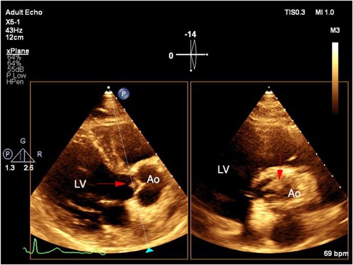
The latest issue of CASE is now available with intriguing reports, including “The Complex Hemodynamic Interplay between Mitral Arcade, Midventricular Obstruction, and Prosthetic Aortic Valve” by Taverna et al. CASE Editor-in-Chief Vincent Sorrell, MD, FASE, remarked, “One of the great values of CASE is the ability to use high-quality echo images matched to tomographic cardiovascular imaging to educate readers about rare diseases they are unlikely to see in their individual clinical practice. This month’s issue fulfills that goal with the report from Taverna that showcases mitral arcade; there are not very many published reports in adults owing to both its rarity and possible under-diagnosis. During the editorial review process, these authors worked closely with section editors who are experts in congenital heart disease to provide readers with very precise descriptions, concluding that this patient likely met criteria for a partial Shone’s complex. They also use very detailed volume-rendered 3D-CT images to demonstrate how the pathoanatomy of this particular mitral arcade is atypical and consists of a double-bridge connecting the anterior and posterior leaflets to the papillary muscles. In summary, the authors rightly conclude that a comprehensive multimodal cardiac imaging approach (CT for anatomy; echo Doppler for physiology) is essential!”
Following that Multimodality Imaging report, authors in Infections in the Heart present a report titled, “Vegetations Lurking in the Dark and the Role of Neoendothelialization,” reviewing the published literature on this potential complication and discussing the possible risk-assessment role of cardiac CT by monitoring neoendothelization in these devices. Over in the Hemodynamic Corner of CASE, there are two reports: one on the importance of spectral Doppler in detecting mitral regurgitation in patients with acoustic shadowing from prosthetic valves, and one that describes the unique mitral and tricuspid spectral Doppler inflow patterns in a patient with non-sinus rhythm. With high-quality 2D and 3D echo images, a case in Rare But Deadly Findings offers a learning lesson to readers about a left atrial dissection after complications of mitral valve surgery. To round out this issue, there is a letter to the editor discussing a previous CASE on a novel transcatheter approach to treat primum atrial septal defects with a response from that article’s authors.
As with all CASE issues, these reports demonstrate the value of quality imaging over quantity. Be sure to read Dr. Sorrell’s editorial as he explores how to maintain this principle in a healthcare environment that increasingly rewards growth and productivity.
Looking for a journal to submit your case report to? We want to hear from you! Email us with questions or submit your report today.
Arthur Weyman ASE’s 8th President Passed Away
It is with great sadness that we write to inform our members that Arthur (Ned) E. Weyman, MD, FASE, passed away this morning, June 17, 2024. Dr. Weyman was ASE’s first secretary of the Board of Directors and subsequently served as the 8th President of ASE serving from January 1991 through June 1993. He received a 2001 American College of Cardiology Gifted Teacher Award and a University of Medicine and Dentistry of New Jersey 2004 Distinguished Alumnus Award. ASE’s Arthur E. Weyman Young Investigator’s Award (YIA) was created in 2007 in honor of Dr. Weyman’s unwavering commitment to research in cardiovascular ultrasound. He received ASE’s Meritorious Service Award in 2012 recognizing his contributions to the field and ASE. Dr. Weyman was also a driving force behind the creation of the National Board of Echocardiography. He served as their first president and remained on their board in an emeritus status. In his honor, the NBE has supported the ASE’s YIA award since 2007.
He completed his medical degree at New Jersey College of Medicine in 1966 and proceeded to his first residency at St. Vincent’s Hospital in New York. He then put his residency on hold while he served in the U.S. Navy as a Marine squadron and air group flight surgeon from 1968-1971. Upon returning, he completed a second year and chief residency at St. Vincent’s, followed by a three-year cardiology fellowship at Indiana University, where he was a fellow in training under ASE’s Founder Dr. Harvey Feigenbaum. In 1980, Dr. Weyman moved to Massachusetts General Hospital and Harvard Medical School in Boston, MA, where he spent the rest of his illustrious career before retiring in 2022. Named Mass General’s chief of cardiology in 1994, his development of innovative methods, research models and exemplary mentoring led the way for echocardiography to become the go-to assessment tool for heart and vascular imaging. He built the echocardiography laboratory into a powerhouse for research, education, and clinical productivity. Known as “the mentor of mentors,” Dr. Weyman and the Mass General echo lab became the place where countless physicians and sonographers competed for fellowships to train in his lab and learn to be great echocardiographers and leaders including seven ASE Presidents.
As one of the founding leaders in echocardiography, he advocated tirelessly for international recognition of the pivotal role the noninvasive assessment tool plays in saving lives by quickly detecting cardiac issues in patients from infants to adults. His presence at the helm of ASE and NBE changed the field forever. He will be missed and we are forever grateful for his service.
A devoted family man, Weyman leaves behind his wife, Jean, their four children and grandchildren.
Listen to a 2009 interview with Dr. Weyman on ASE’s YouTube channel.
A funeral Mass for Dr. Weyman will be held in St. Paul Church, 502 Washington St., Wellesley on Saturday, June 22, at 10AM. Relatives and friends kindly invited. The Mass will also be live streamed at www.harborview.live. Interment in Woodlawn Cemetery, Wellesley.
ASE 2024 Research Spotlight 4
FOR IMMEDIATE RELEASE
Contact: Angie Porter
919-297-7152
APorter@ASEcho.org
Echocardiography Plays an Important Role in Identifying Risk of Future Hypertension in Preeclampsia Patients
(DURHAM, NC, June 14, 2024)—Research that will be presented at the American Society of Echocardiography’s 35th Annual Scientific Sessions, June 14-16, 2024, in Portland, Oregon, will share how echocardiography can be used to help predict the future development of hypertension in women who had preeclampsia during pregnancy.
Preeclampsia, a condition characterized by high blood pressure and organ damage during pregnancy, affects numerous women worldwide. After giving birth, some women who experienced preeclampsia are at an increased risk of developing hypertension. However, accurately identifying those at risk can be challenging, and there is currently limited research on how echocardiographic changes influence outcomes.
The research study, titled “Echocardiographic Parameters Associated with Future Hypertension in Patients with Preeclampsia,” intended to address that knowledge gap by utilizing echocardiography performed in the first three months post-preeclampsia to identify specific findings that could indicate an increased risk of high blood pressure in the future.
Researchers retrospectively examined echocardiographic predictors, such as left ventricular mass index (LVMi) and increased interventricular septal thickness (IVSD), as parameters for future diagnosis of hypertension. Out of the 252 women included in the study, which was conducted at Allegheny Health Network in Pittsburgh, Penn., nearly 50 percent were subsequently diagnosed with hypertension during follow-up within 2.3 years.
“Our study used echocardiography to check for specific findings that could identify women who might be at higher risk for high blood pressure after preeclampsia,” said lead author Dr. Anushree Puttur, a resident physician at the Allegheny Health Network. “By finding these clues, we hope to improve how we monitor and care for women after they’ve had preeclampsia.”
ASE 2024 will feature more than 450 abstract presentations showcasing cutting-edge research on the latest advances in cardiovascular ultrasound and cases illustrating breakthroughs in patient care. This research study will be presented as a rapid-fire abstract presentation on Saturday, June 15, from 4:00 to 5:00 p.m. at the Oregon Convention Center. Learn more about ASE 2024 and download the conference’s final event program at ASEScientificSessions.org.
About American Society of Echocardiography
The American Society of Echocardiography (ASE) is the Society for Cardiovascular Ultrasound Professionals™. ASE is the largest global organization for cardiovascular ultrasound imaging serving physicians, sonographers, nurses, veterinarians, and scientists and as such is the leader and advocate, setting practice standards and guidelines for the field. The Society is committed to advancing cardiovascular ultrasound to improve lives. For more information visit ASE’s website ASEcho.org or social media pages on Facebook, X (formerly Twitter), LinkedIn, or Instagram.
###
ASE 2024 Research Spotlight 3
FOR IMMEDIATE RELEASE
Contact: Angie Porter
919-297-7152
APorter@ASEcho.org
Artificial Intelligence in Cardiovascular Ultrasound Imaging Research Highlighted at ASE 2024
(DURHAM, NC, June 14, 2024)—Artificial intelligence (AI) continues to transform the field of cardiovascular ultrasound imaging, and AI-focused research is a prominent topic at the American Society of Echocardiography’s 35th Annual Scientific Sessions, taking place June 14-16, 2024, in Portland, Oregon.
This year’s meeting is aptly named Innovations in Diagnosis and Beyond and will feature more than 450 abstract presentations showcasing cutting-edge research on the latest advances in cardiovascular ultrasound and cases illustrating breakthroughs in patient care.
Two research studies being presented on Saturday, June 15, examined a Food and Drug Administration (FDA)-cleared AI tool with the potential to streamline echocardiography exams and better identify patients with heart failure with preserved ejection fraction (HFpEF).
The first research study, titled “Real-World Clinical Validation of an Artificial Intelligence Pipeline for Automated Assessment of LV Function,” investigated the potential of AI-guided software in accurately and consistently measuring left ventricular ejection fraction (LVEF) and global longitudinal strain (GLS), which are crucial in the diagnosis and management in patients with HFpEF.
Current practices often require sonographers to acquire multiple examples of the same view of the heart during an echocardiography exam and then retrospectively measure the most optimal image. This can lead to increased exam times, musculoskeletal injuries, and measurement variabilities. To address these challenges, researchers used AI technology to examine nearly 150 clinical echocardiograms and assess the effectiveness of an automated AI-driven process in providing reliable measurements.
Research findings indicated that an AI algorithm that automatically identifies images and provides GLS and LVEF in HFpEF patients may be a valuable tool in addressing sonographer and physician workflow, but also emphasized the need for further efforts to increase agreement between manual and automated echocardiography measurements.
“The echocardiography exam workflow presents a promising opportunity for AI to streamline processes and improve patient diagnoses,” said the study’s lead author Madeline Jankowski, BS, RDCS, ACS, FASE, a cardiac sonographer and clinical research associate at Northwestern University in Chicago. “At Northwestern Medicine, our collaboration involves a multidisciplinary team of physician and sonographer experts who evaluate emerging AI software for its diagnostic capabilities and utility within a busy healthcare institution. Given the complexity of incorporating AI into echocardiography, it remains crucial to include all stakeholders in ongoing discussions to address workflow and quality issues effectively.”
In the second study, titled “AI-Based Detection of Heart Failure with Preserved Ejection Fraction,” researchers also aimed to test the capabilities of AI technology by evaluating nearly 700 clinical echocardiograms during a seven-day period. The novel AI software used a single four-chamber apical transthoracic echocardiogram to identify patients with HFpEF.
While the findings emphasized the need for additional testing to clarify diagnoses, especially in cases where certain parameters may impact the precision of existing tools, they also demonstrated comparable accuracy to established diagnostic tools in recognizing patients necessitating further evaluation for HFpEF. The AI software helped identify 68 percent more potential cases of HFpEF in patients undergoing routine echocardiograms.
“HFpEF is the most prevalent form of heart failure globally. However, pinpointing it can be difficult due to its varying presentation in patients,” said the study’s lead author Amogh Karnik, MD, MSc, a cardiovascular disease fellow at the McGaw Medical Center at Northwestern University. “In this study, we assessed a novel AI tool designed to detect signs of HFpEF by analyzing ultrasound images of the heart. Such a tool holds promise for early diagnosis of HFpEF, and can help facilitate earlier initiation of treatment.”
Learn more about ASE 2024 and download the conference’s final event program at ASEScientificSessions.org.
About American Society of Echocardiography
The American Society of Echocardiography (ASE) is the Society for Cardiovascular Ultrasound Professionals™. ASE is the largest global organization for cardiovascular ultrasound imaging serving physicians, sonographers, nurses, veterinarians, and scientists and as such is the leader and advocate, setting practice standards and guidelines for the field. The Society is committed to advancing cardiovascular ultrasound to improve lives. For more information visit ASE’s website ASEcho.org or social media pages on Facebook, X (formerly Twitter), LinkedIn, or Instagram.
###
ASE 2024 Research Spotlight 2
FOR IMMEDIATE RELEASE
Contact: Angie Porter
919-297-7152
APorter@ASEcho.org
Exploring Innovative Applications of Artificial Intelligence in Echocardiography at ASE 2024
(DURHAM, NC, June 14, 2024)—Echocardiography plays a foundational role in evaluating individuals with suspected cardiac conditions, such as hypertrophic cardiomyopathy (HCM) and cardiac amyloidosis (CA). Both HCM and CA can be difficult to distinguish in clinical practice, so researchers are examining the potential benefits of augmenting echocardiography with artificial intelligence (AI) to benefit patients in these cohorts.
The findings from two research studies exploring the application of AI with echocardiography in accurately diagnosing and managing HCM and CA patients will be presented as abstract poster presentations during the American Society of Echocardiography’s 35th Annual Scientific Sessions, June 14-16, 2024, in Portland, Oregon.
Utilizing Machine Learning for More Accurate and Expedited Pediatric HCM Diagnosis
HCM can be present at any age and difficult detect since patients are often asymptomatic. Moreover, HCM is a significant cause of sudden cardiac death in children, making early and accurate detection necessary.
The research study titled, “Identification of HCM in Children by Machine Learning Applied to Echocardiography,” developed machine learning models that provide accurate and expedited HCM diagnosis in children. The machine learning models created for this research study analyzed datasets from more than 30 years of echocardiography data of HCM patients. The information generated from these models will help guide medical teams in formulating patient treatment plans.
“Echocardiography is the imaging modality considered to be the gold standard for HCM diagnosis. Our research introduces an innovative application of AI to enhance pediatric HCM diagnosis through the automated analysis of echocardiography data in real-time,” says the study’s lead author Vanessa Karlinski Vizentin, MD, a research fellow at Mayo Clinic.
Mayo Clinic Cardiologist Adelaide M. Arruda-Olson, MD, PhD, who is a senior author of the study added, “This forward-thinking approach holds immense promise for optimizing healthcare delivery, ensuring that children affected by HCM receive timely diagnosis, interventions, and personalized care.”
AI with Echocardiography May Prevent Diagnostic Delays, Better Detect CA
CA is a disorder in which abnormally folded proteins deposit in the heart resulting in heart failure. CA is considered a rare disease in older adults; however, recent studies have shown that CA may be more common than initially thought.
A research study titled, “Novel Deep Learning Model for the Detection of Cardiac Amyloidosis: A Multicenter, International Study” tested AI’s ability to improve the detection of CA with echocardiography, allowing for earlier diagnosis and the initiation of life-saving therapies. The study’s deep learning model was able to accurately differentiate CA from phenotypic controls with increased wall thickness. This finding may augment the screening accuracy of echocardiography, preventing diagnostic delays and helping to better identify individuals at the highest risk for CA.
“Although echocardiography is frequently the first cardiac imaging test ordered to detect cardiac amyloidosis, classical echocardiographic features may miss many patients,” says the study’s lead author Jeremy Slivnick, MD, FASE, an assistant professor at The University of Chicago Medicine. “In our study, we developed a novel AI model which could accurately detect CA using only an apical four-chamber view. Our AI model has the potential to improve the echocardiographic detection of CA, allowing for earlier diagnosis and treatment of this life-threatening disorder.”
ASE 2024 will feature more than 450 abstract presentations showcasing cutting-edge research on the latest advances in cardiovascular ultrasound and cases illustrating breakthroughs in patient care. Both of these AI research studies will be presented as poster presentations on Saturday, June 15, from 12 to 12:45 p.m. at the Oregon Convention Center. Learn more about ASE 2024 and download the conference’s final event program at ASEScientificSessions.org.
About American Society of Echocardiography
The American Society of Echocardiography (ASE) is the Society for Cardiovascular Ultrasound Professionals™. ASE is the largest global organization for cardiovascular ultrasound imaging serving physicians, sonographers, nurses, veterinarians, and scientists and as such is the leader and advocate, setting practice standards and guidelines for the field. The Society is committed to advancing cardiovascular ultrasound to improve lives. For more information visit ASE’s website ASEcho.org or social media pages on Facebook, X (formerly Twitter), LinkedIn, or Instagram.
###
ASE 2024 Research Spotlight 1
FOR IMMEDIATE RELEASE
Contact: Angie Porter
919-297-7152
APorter@ASEcho.org
Two Research Studies at ASE 2024 Examine Environmental Factors and Heart Health
(DURHAM, NC, June 14, 2024)—A number of factors can determine a person’s heart health—genetics, physical activity, diet, and even the environment. Two international research studies that will be presented during the American Society of Echocardiography’s (ASE) 35th Annual Scientific Sessions, June 14-16, 2024, in Portland, Oregon, examined the effects the latter on the heart.
The first study, titled “Maternal Exposure to Ambient Ozone and Fetal Conotruncal Congenital Heart Defects in China: A Large Multicenter Cohort Study,” delves into the effects of ozone (O3) pollution on fetal heart development. From January 2013 to December 2021, the study examined more than 24,000 pregnant women at Beijing Anzhen Hospital, Capital Medical University in China to investigate the association between maternal exposure to O3 and conotruncal heart defects (CTDs), which are congenital heart malformations that affect the heart’s outflow tracts and great arteries.
Fetal echocardiography was used to evaluate fetuses with CTDs, and the research findings revealed that maternal exposure to ambient O3 pollution during critical periods, especially during the periconception period, may increase the risk of CTDs in fetuses.
“To prevent this impact, pregnant women should adopt healthy lifestyles and avoid exposure to O3 pollution during peak hours, which can occur in traffic congested or industrial areas,” says the study’s lead author, Yihua He, an expert in fetal congenital heart disease and the Director of the Echocardiography Medical Center and Maternal-fetal Medicine Center in Fetal Heart Disease at Beijing Anzhen Hospital, Capital Medical University. “Additionally, governments and the public should implement measures to decrease O3 pollution, such as reducing emissions from industrial and transportation sources, promoting clean energy, and implementing air quality standards.”
Dr. He emphasized the need for further research to understand the impact of other environmental factors on fetal heart development and to develop appropriate preventive measures.
The second study, titled “Echocardiographic Assessment of Pulmonary and Right Chamber Parameters in Healthy Individuals Who Live More Than 8200 Feet Above the Sea Level: A Colombian Experience,” focused on long-term cardiopulmonary adjustments in individuals residing at high altitudes, specifically those living more than 8,200 feet above sea level (FASL).
The study, conducted in the Andean Mountains in Colombia, where over 35 million people live at such altitudes, examined the prevalence of high-altitude pulmonary hypertension and its implications on the right ventricle and clinical outcomes. Contrary to what was expected, the findings indicated that individuals living above 8200 FASL do not exhibit higher estimated systolic pulmonary artery pressure (sPAP) or significant differences in right ventricle dimensions and function.
However, findings did discover a significant difference between the study’s two Colombian cohorts from Bogota (8,612 FASL) and Aquitania (9,941 FASL). There was a surprisingly higher prevalence of Stage 2 hypertension and an increase in relative wall myocardium thickness in the Aquitania cohort. It was determined there is a need for further investigation into the long-term adaptive mechanisms in adults residing at high altitudes, as it may lead to adverse cardiovascular outcomes.
“The effects of pulmonary and arterial hypertension include capillary remodeling and permeability changes that result in pressure load on the right or left ventricle respectively. If uncontrolled, changes can lead to ventricular failure with a reduced exercise capacity and premature death. Therefore, if undiagnosed, high-altitude dwellers could be at risk of developing clinically relevant events,” says the study’s lead author Astrid Lorena Rodriguez, an internal medicine resident at Fundacion Santa Fe, Bogota in Colombia. “We expect our results to encourage global leaders and colleagues to further research long-term cardiopulmonary adjustments to better understand clinical outcomes in this population.”
Both research studies will be presented at the Oregon Convention Center during ASE 2024, which features more than 450 abstract presentations showcasing cutting-edge research on the latest advances in cardiovascular ultrasound and cases illustrating breakthroughs in patient care. Learn more about ASE 2024 and download the conference’s final event program at ASEScientificSessions.org.
About American Society of Echocardiography
The American Society of Echocardiography (ASE) is the Society for Cardiovascular Ultrasound Professionals™. ASE is the largest global organization for cardiovascular ultrasound imaging serving physicians, sonographers, nurses, veterinarians, and scientists and as such is the leader and advocate, setting practice standards and guidelines for the field. The Society is committed to advancing cardiovascular ultrasound to improve lives. For more information visit ASE’s website ASEcho.org or social media pages on Facebook, X (formerly Twitter), LinkedIn, or Instagram.
###
Revolutionary Cardiovascular Ultrasound Innovations Being Showcased at ASE 2024 in Portland, Oregon
FOR IMMEDIATE RELEASE
Contact: Angie Porter
919-297-7152
APorter@ASEcho.org
Revolutionary Cardiovascular Ultrasound Innovations Being Showcased at ASE’s 35th Annual Scientific Sessions in Portland, Oregon
(PORTLAND, Ore., June 11, 2024) — The future of cardiovascular ultrasound is being showcased at the American Society of Echocardiography (ASE) 35th Annual Scientific Sessions, June 14-16, 2024, at the Oregon Convention Center in Portland, Oregon. From cutting-edge cloud-based imaging technology to simulators and FDA-cleared devices, the innovations being highlighted will lead to more accurate diagnoses and personalized patient treatment.
Fifty companies and organizations will display their latest products and services, including ICE catheter technologies, software systems for integration with medical records, and solutions for creating a safe and ergonomic work environment. ASE 2024 is the world’s premier meeting for cardiovascular ultrasound practitioners and promises a wealth of education, research and the latest vendor technology. The Echo Expo will be open Friday, June 14, 3:30 – 5:30 p.m.; Saturday, June 15, 10 a.m. – 5:30 p.m. and Sunday, June 16, 10 a.m. – 2 p.m.
Highlights from the show floor, as reported in the vendors’ own words, will include:
ASE (Booth 654) will be selling both physical and online educational products at discounted prices. This year, ASE’s ImageGuideEcho™ will share a space in the ASE booth. There will be many opportunities in the ASE booth for attendees to talk with leadership on important topics, like how to achieve the Fellow of the American Society of Echocardiography (FASE) designation, become a volunteer, and apply for ASE’s Leadership Academy Cohort 4.
The ASE Foundation (ASEF) Booth, located in the Pre-Function C Lobby on Level 1, is the central hub for all the Foundation’s charitable activities and initiatives. Attendees are encouraged to join the Cardiovascular Challenge, sponsored by Cardiovascular Credentialing International, to promote moving and cardiovascular health. There is also a special showcase of photographs called Images from the Heart. The Foundation has awarded scholarships and travel grants for this year’s Scientific Sessions to 75 early career practitioners, totaling over $96,000 in support.
Abbott (Booth 431) will showcase its recently U.S.-approved TriClip G4 Transcatheter Edge-to-Edge Repair (TEER) system. With maximized effectiveness, exceptional safety and life-changing impact, this innovative device is redefining what is possible in tricuspid treatment. Visit Abbott to learn more about how you can help more of your patients suffering from symptomatic, severe tricuspid regurgitation enjoy a quality of life they never thought possible!
AGFA HealthCare (Booth 247) will present its groundbreaking Enterprise Imaging solution, tailored for cardiology, and providing unparalleled support for the entire cardiology department across all its specialties. With its comprehensive web workflows, cardiologists can achieve a “live in flow” – conveniently access their images and reports anytime and anywhere, resulting in a revolutionary Imaging Health RecordTM that prioritizes what’s most important – patient care. This innovative solution seamlessly integrates with the hospital’s medical record system, allowing for collaboration and enhanced efficiency in delivering exceptional cardiac care.
AstraZeneca (Booth 550) will drive brand awareness and excitement to promote WAINUA as the silencer of choice in patients with polyneuropathy due to hereditary amyloidosis (hATTR-PN). The booth will feature the WAINUA HCP Campaign to Stop Neuropathy Progression and will include various additional informational resources.
Bristol Myers Squibb (Booth 855) continues to be an innovator in cardiology. Our mission to deliver for patients is what drives us. This year at ASE, we focus on the symptomatic oHCM patient and how our therapy potentially offers patients more. Join us and explore what current patients are experiencing.
CHRISTUS Health (Booth 343) Recruitment of Ecotech positions is a pioneering entity within the healthcare industry, dedicated to sourcing and employing top-tier Ecotechs for integration within our healthcare facilities. Our mission is twofold: to ensure the provision of high-quality healthcare services while simultaneously championing environmental sustainability and eco-conscious practices within our operations. Stop by and speak with us about your future!
Dell Children’s Medical Center (Booth 434) The Texas Center for Pediatric and Congenital Heart Disease is a clinical partnership between Dell Children’s Medical Center and UT Health Austin. Our multidisciplinary, multispecialty teams bring their expertise together to ensure delivery of the highest level of individualized and comprehensive care possible to even the tiniest most complex patients, as well as adults with congenital heart disease, for best possible outcomes. Visit us to learn more.
EchoNous (Booth 334) is at the forefront of point-of-care ultrasound (POCUS) innovation, revolutionizing the field by introducing an all-new POCUS category, powered by AI. With Kosmos, users get both exceptional image quality and advanced performance of cart-based systems, with the affordability and unmatched portability of a handheld system. Layer on top of that industry-leading AI that helps flatten the learning curve and makes exam workflows more efficient. Now you have a POCUS solution that is both accessible for new users and powerful enough for experts.
Epsilon Imaging (Booth 650) EchoInsight® Zero Footprint (ZF) Advanced Echo Analysis is an integrated, vendor neutral, offline platform, enabling clinicians to easily incorporate the valuable benefits of strain imaging and automation of cardiac function measurements into routine clinical practice. EchoInsight ZF offers robust yet economical packages to accommodate a variety of customer needs: LV and RV analysis, full heart analysis and/or clinical research analysis capabilities. Applications are based on ASE guidelines and recommendations, and scale to meet customer workflow needs.
Fujifilm Healthcare Americas Corporation (Booth 435) Visit to see the transformative potential of our diagnostic ultrasound and enterprise imaging innovations to inspire the imaging techniques of tomorrow.
HeartLab (Booth 530) will be demoing its cloud-based cardiology imaging platform, which is becoming increasingly popular with cardiac care teams around the world. Replacing outdated PACS, HeartLab offers productivity and efficiency gains, and the ability to work with remote team members, improving the level of patient care. HeartLab has also expanded its off-cart measurement suite to ensure that almost all custom calculations can now be performed in the cloud without having to return to the machine.
Heritage Medical Products (Booth 551) aka The SONOBED Company. The bestselling cardiac exam bed since 2001, the SonoBed ATLAS, from Heritage Medical Products Inc. will be shown in the booth. The Atlas made its first debut at the ASE Scientific Sessions in 1997, and has continued to support ASE by attending every year. Stop by to learn more!
ImageGuideEcho Registry (Booth 654) The ImageGuideEcho Registry™ is an echocardiography data repository for quality improvement owned and operated by ASE. The ImageGuideEcho Registry provides automatically generated QI feedback and analytics for the benefit of stakeholders, including patients, physicians, and researchers. The Registry focuses on driving technology applications and assessing specific quality metrics and patient outcomes across medical centers within the field of echocardiography. There will be two information sessions in the Registry Booth this year. Visit booth 654 to learn how you and your colleagues can be a part of the growing Registry for the benefit of patients, your institution, and the field of echo.
Intersocietal Accreditation Commission (IAC) (Booth 436) IAC Echocardiography’s two latest offerings provide opportunities to demonstrate a commitment to quality and patient safety in specialized diagnostic imaging:
- The IAC Standards & Guidelines for Perioperative Transesophageal Echocardiography provide guidance in training and experience, protocol development and resources.
- Adult Congenital Transthoracic Echocardiography Accreditation recognizes the critical role of facilities providing care to patients with congenital heart disease who transition their care from pediatric cardiology to adult cardiology services.
Joint Review Committee on Education in Diagnostic Medical Sonography (JRC-DMS) (Booth 748) is a nonprofit organization in existence to establish, maintain and promote quality standards for educational programs in Diagnostic Medical Sonography (DMS). Since 1983, the organization has provided a mechanism of committee review to recognize educational programs throughout the United States that instruct students in the disciplines related to DMS through education consistent with standards for entry into practice.
Lantheus (Booth 755) The safety and effectiveness of DEFINITY® have been established for use in pediatric patients with suboptimal echocardiograms to opacify the left ventricular chamber and to improve delineation of the left ventricular endocardial border. Use of DEFINITY® for this indication is supported by evidence from adequate and well-controlled studies in patients starting at one month of age. Come visit our booth to learn more and enjoy a latte!
Mayo Clinic (Booth 449) Cardiovascular Education department is showcasing their 2024/2025 lineup of live conferences at world-class destinations, as well as new and exciting online digital learning opportunities. Stop by to interact with top physicians and sonographers and learn how you can connect and collaborate with Mayo Clinic in research, education and patient care.
narnar (Booth 651) Our mission at narnar is to enable the widespread use of contrast echocardiography in diagnosing heart disease by developing clinical software tools that make the interpretation of myocardial function and perfusion faster, consistent and more intuitive. Please visit us at our booth to learn more!
National Board of Echocardiography (NBE) (Booth 750) launches its new Maintenance of Certification in Echocardiography (MOCE) this July 2024. Come by our booth for a MOCE platform demo, on a mobile device or web browser. NBE Testamurs and Diplomates will begin an annual cycle of answering questions quarterly at their own pace. This annual longitudinal assessment replaces NBE recertification exams to continuously support physicians in keeping their clinical knowledge, judgment, and skills current throughout their careers.
NeuraSignal (Booth 350) is pleased to showcase the NeuraSignal NovaGuide 2 Intelligent Ultrasound, a robot-assisted transcranial Doppler (TCD) device that automates the detection of right-to-left shunts (RLS) and patent foramen ovale (PFO). The BUBL study demonstrated that this device is significantly superior in sensitivity compared to transthoracic echocardiography (TTE), with a 64 percent RLS detection rate by NovaGuide 2 versus only 20 percent by TTE, highlighting its enhanced accuracy and potential for broader clinical use. We are excited to demonstrate its full capabilities.
Philips (Booth 441) welcomes you to the next dimension in echo, introducing new AI tools like the first automated tool for Segmental Wall Motion scoring of the heart and the first fully automated 3D quantification of MR volumes* to speed workflows and increase productivity. Plus, see our innovative X11-4t Mini 3D TEE transducer which provides care to patients as small as 5kg and beyond. *Pending 510(k) clearance.
Pie Medical Imaging (PMI) (Booth 337), a global leader in cardiac imaging, showcases their innovative echocardiography solutions. Caas Qardia leverages FDA-cleared artificial intelligence driven workflows for performing key clinical measurements including myocardial strain, stress echo, diastology and valve analysis. Ease-of-access is provided on any PC within the hospital network by using the web-browser. Also, PMI presents their CT/3DTEE-based pre-procedural transcatheter planning solutions within the 3mensio Structural Heart platform.
ScImage (Booth 744) PICOM365 comprehensive CVIS continues to move ahead with specific echocardiology advancements. Expanded structured reporting optimization leveraging AI, along with continuous efficiency enhancements, ensure a state-of-the-art offering. Our cloud-native solution allows anytime, anywhere reading, meeting all viewing, reporting, and data distribution needs, and supports a full complement of current AI third-party systems from any location. Discover what our echocardiogram system offers.
Sheehan Medical (Booth 645) the Gold Standard in ultrasound education. We give you the keys to make ultrasound easy to learn and easy to teach:
- Instant feedback that shows you how you’re doing AND how to improve further.
- Auto import so you can upload your best patient images onto the simulator.
- Our latest innovation: CT as well as real 3D echo images on our TEE simulator.
Siemens Healthineers (Booth 741) boasts 5600+ AI-powered measurements revolutionizing cardiac care. Discover ACUSON Origin’s suite of AI tools with the latest heart simulator for hands-on TEE and 4D ICE training.
SoftLink International (Booth 351) is a health-tech company founded in 1997. It provides IT solutions for hospital automation, clinical informatics, patient workflow management and analytics. SoftLink is excited to showcase its Comprehensive Echocardiography Solution. The solution comprises an Adult Echocardiography Procedure Reporting System and a Diagnostic Ultrasound Viewer within an EchoPACS – a scalable Image Management environment. It supports on-premise and cloud deployment options with modality integration for the auto-import of DICOM measurements.
Ultromics (Booth 849) will show insights into the latest innovations in heart failure detection with demos of the unique AI-powered, echo-based diagnostic technology. Don’t miss this chance to see how Ultromics is revolutionizing cardiac care!
UCSF Health (Booth 257) Join our world class organization. At UCSF Health, you’ll work with a team of talented clinical sonographers, ACHD sonographers, advanced cardiac sonographers, research sonographers, core lab sonographers and echo students from our hospital-based cardiac sonography school, all supported by a dedicated full time sonographer educator and a sonographer leadership team. Meet our UCSF Health team to learn more about our opportunities!
UVA Health (Booth 430) is a world-class academic medical center and health system with a level 1 trauma center. Its flagship hospital, UVA Health University Medical Center, is ranked #1 in Virginia and is among the top 40 nationally by Newsweek’s “World’s Best Hospitals 2024” guide. Our ECHO lab received the Silver Anniversary award for 25 years of commitment to quality. Visit us and find your future with UVA Health.
Valley Children’s Healthcare (Booth 354) Pediatric Cardiology Department offers cardiac testing services for children and mothers expecting, providing comprehensive care for heart conditions. This state-of-the-art center provides a wide range of testing services to patients of all ages, ensuring accurate diagnoses and effective treatment plans are developed. The focus of the department is to offer collaborative care, utilizing the latest advancements in technology to ensure the best possible outcomes for our patients.
Version2 VR Technologies (Booth 346) announces the launch of its echocardiography simulator. This immersive VR toolkit delivers unparalleled portability, engagement, and realism. Facilitating hands-on learning, the simulator ensures training quality and accessibility. With features such as video guides, cut plane visualization, and transparency mode, users can effectively enhance their skills, confidence, and comprehension. We’re excited to offer a solution that empowers students and healthcare professionals to elevate their expertise in a dynamic and immersive learning environment.
Visura Technologies (Booth 740) is the manufacturer of the TEECAD System, the only FDA-cleared camera that seamlessly connects to a TEE probe, is excited to be showcasing our newest innovations for real-time visualization. Our TEECAD Innovation Station will return to provide ASE attendees a hands-on experience, highlighting the importance of real-time visualization during TEE probe navigation to support more efficient and effective probe placement. Stop by to learn more about this latest technology and available data.
About ASE
The American Society of Echocardiography (ASE) is the Society for Cardiovascular Ultrasound Professionals™. ASE is the largest global organization for cardiovascular ultrasound imaging serving physicians, sonographers, nurses, veterinarians, and scientists and as such is the leader and advocate, setting practice standards and guidelines for the field. The Society is committed to advancing cardiovascular ultrasound to improve lives. For more information visit ASE’s website ASEcho.org or social media pages on Facebook, X (formerly Twitter), LinkedIn, or Instagram. For more information about the ASE Scientific Sessions visit ASEScientificSessions.org.
###
Calling All Future Leaders: ASE Leadership Academy Cohort 4 Application is NOW OPEN
Jumpin’ for June JASE
The June issue of JASE includes, “Obstruction in Hypertrophic Cardiomyopathy: Many Faces.” Author Jeffrey B. Geske, MD, FASE, remarks, “Echocardiographic assessment of obstructive hypertrophic cardiomyopathy can be challenging due to vast phenotypic heterogeneity, complex hemodynamics, and variable findings dependent on loading conditions. However, accurate diagnostic assessment is needed for guidance of medical management, determination of septal reduction therapy candidacy, and intraprocedural assessment. In this JASE article, we present a visual Doppler atlas with many high-quality examples to guide interpretation and understanding of the many ways that obstruction can present in hypertrophic cardiomyopathy.”
In addition to the HCM report, readers can look forward to two other state-of-the-art reviews. Both center on the role of 3D echocardiography—in the perioperative assessment of the hemodynamic ventriculoarterial junction of the aortic root and in RV volume analysis in pediatric heart disease. This issue also hosts a variety of clinical investigations on topics ranging from left ventricular trabeculations in athletes to 3D echocardiography’s role in mitral regurgitation to congenital heart disease. Two editorial comments, five brief research communications, and two letters to the editor round out this issue.
In his final President’s Message, Dr. Eidem reflects on this past year as ASE President and the progress of his three key initiatives: ASE Matters, ASE Mentors, and ASE Educates. Be sure to check out June’s Author Spotlight to learn more from author Giuseppe Di Gioia, MD, on his recent paper, Left Ventricular Trabeculation: Arrhythmogenic and Clinical Significance in Elite Athletes.
Please see the June ASE Education Calendar for a listing of educational opportunities far and wide.