A Standout September CASE

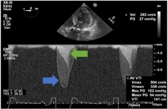
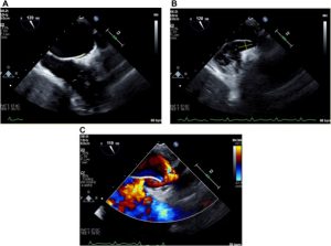
The latest issue of CASE is now available with intriguing reports, including “Multidirectional Blood Flow During Cardiopulmonary Bypass Mimicking an Iatrogenic Aortic Dissection During Transesophageal Echocardiographic Examination.” CASE Editor-in-Chief Vincent Sorrell, MD, FASE, remarked, “Foster et al. remind us all of one of the very important reasons this Journal has been such a success to our Society. These authors elegantly describe a common finding during cardiopulmonary bypass that may result in potentially devastating clinical consequences when not recognized. They performed a series of basic scientific steps to solve the root cause for understanding an image that initially appeared to be an aortic dissection. In doing so, they describe the pattern of multidirectional blood flow within the aorta that accompanies bypass cannulation. Next, they demonstrate the change in Doppler findings when the bypass flow is held. Lastly, they include post-operative tomographic imaging for further education. Readers unfamiliar with this finding may be spared an unnecessary poor outcome. Other readers may use this approach as a guide to their own CASE report when they are confronted with an echo/Doppler artifact.”

A second Intraoperative Echocardiography report follows the sequence of intraoperative clinical events that resulted in severe hypoxemia during aortic valve surgery, using TEE as a primary investigative tool. In the Congenital Heart Disease category, Fahim et al. expound upon their echo findings in an adult with a double-chambered right ventricle, including excellent correlative CMR images to enhance their echo insights. The next case highlights the critical value of peripheral ultrasound with a patient whose pseudoaneurysm found on vascular ultrasound uniquely had a second pseudoaneurysm evolving from the first. Just Another Day in the Echo Lab rounds out this issue with a reminder that serial TEE is crucial after placement of a left atrial appendage occlusion device.

Be sure to read Dr. Sorrell’s editorial as he shares insights from patients on how to better engage them in the echo process, so they can feel like informed, active participants in their progression of care.

Looking for a journal to submit your case report to? We want to hear from you!
Email us with questions or submit your report today.

Register Early for SOTA and Save!

For nearly four decades, ASE’s State-of-the-Art Echocardiography™ (SOTA) has featured outstanding faculty who share the latest in echocardiography education with attendees. The 37th Annual SOTA will take place February 14-17, 2025, at the Westin Kierland Resort & Spa in Scottsdale, Arizona. The 2025 program follows the themes of structural heart disease, myocardial and pericardial disease, coronary artery disease, and interventional echocardiography. During the course, presentations will highlight what echocardiography brings to each theme, help clinicians recognize and avoid common errors and pitfalls, and demonstrate how to apply the guidelines in a practical manner. Additionally, participants who also sign up for the Preconference Learning Lab will have the opportunity to interact with the faculty one-on-one. Register early to save up to $225!

Pledge Your Support for ASEF’s Coffee Day

Get ready for Coffee Day 2024! Mark your calendars from September 29 – October 5 to help the ASE Foundation raise $5,000 to support our newest initiative: Cardiovascular Sonographer Leadership Development. Funding toward this initiative aims to provide support for sonographer members volunteering in leadership positions within ASE and ASEF.

Alan D. Waggoner Student Scholarship Winner Alexandre Campos Dias, RDCS, has already pledged his support. Thank you, Alex! If you’re planning to contribute, join Alex by making your pledge known. Contact the ASE Foundation at Foundation@ASEcho.org.

Want to be an ASE Soulmate?

In 2025, ASE is offering a variety of new membership promotions to celebrate our 50th Anniversary and reach our goal of 20,000 members! One of these new promotions, ASE Soulmates, is replacing the current ASE Ambassador program in 2025. To become an ASE Soulmate, refer two new members to join ASE by August 31, 2025, and save 25% on your 2026 membership. Refer three or more new members and save 50% on your 2026 membership! Some exclusions apply, so be sure to visit the ASE Soulmates page for details.

YOU make ASE the largest voice in cardiovascular imaging in the world and help create a stronger community of collaboration that advances the field and improves patient lives! We thank you for your membership and want to recognize the 137 ASE Ambassadors who participated in 2024 resulting in 353 new members!

ASE Publishes Updated Guideline to Improve Care for HF Patients with Implanted Medical Devices

FOR IMMEDIATE RELEASE

Contact: Angie Porter
919-297-7152
APorter@ASEcho.org

ASE Publishes Updated Guideline to Improve Care for Heart Failure Patients
with Implanted Medical Devices

(DURHAM, NC, September 10, 2024)—A new guideline from the American Society of Echocardiography (ASE) aims to provide more detail on the best imaging strategies for managing and improving care for heart failure patients with surgically implanted left ventricular assist devices (LVADs) and temporary mechanical circulatory support (TMCS) devices.

Advances over the past several years prompted the need for an updated perspective on the role of echocardiography and multimodality imaging to evaluate and manage potential complications associated with temporary and durable LVADs and TMCS devices. Recommendations for Multimodality Imaging of Patients with Left Ventricular Assist Devices and Temporary Mechanical Support: Updated Recommendations from the American Society of Echocardiography is an update to the Society’s original guideline published in 2015.

“This new guideline document serves as an update to the 2015 comprehensive ASE LVAD guideline. We provide detailed information and recommendations on TMCS devices, and we define the contemporary role of echocardiography and multimodality imaging in patient selection, pre-intra-and post-procedural surveillance, and troubleshooting with focus on the HM3 LVAD,” said lead author Dr. Jerry Estep, the Division Chair of Cardiovascular Medicine at Cleveland Clinic Florida in Weston. “We hope the framework we provide will improve patient outcomes by providing the best imaging strategies before and after durable and temporary device implantation.”

Many principles and recommendations detailed in ASE’s original 2015 guideline are still current and valid, and this updated document includes several of the previously published key points for the use of echocardiography. Additionally, it addresses new devices, expands upon the role of multimodality imaging and acknowledges the growing role of point-of-care ultrasound in evaluating patients with mechanical circulatory support. The writing group includes experts from multiple disciplines who hope the document will encourage future studies to continue advancing the field.

The new guideline is published in the September 2024 issue of the Journal of the American Society of Echocardiography. All guidelines published by ASE are available at ASEcho.org/Guidelines.

About American Society of Echocardiography
The American Society of Echocardiography (ASE) is the Society for Cardiovascular Ultrasound Professionals™. ASE is the largest global organization for cardiovascular ultrasound imaging serving physicians, sonographers, nurses, veterinarians, and scientists and as such is the leader and advocate, setting practice standards and guidelines for the field. The Society is committed to advancing cardiovascular ultrasound to improve lives. For more information, visit the ASE website ASEcho.org or social media pages on Facebook, X (formerly Twitter), LinkedIn, or Instagram.

###

New ASE Guideline Clarifies Cardiac POCUS Nomenclature

FOR IMMEDIATE RELEASE

Contact: Angie Porter
919-297-7152
APorter@ASEcho.org

New ASE Guideline Clarifies Cardiac Point-of-Care Ultrasound Nomenclature
The guideline aims to standardize the nomenclature around cardiac point-of-care ultrasound to facilitate collaboration, reduce confusion and enhance patient safety

(DURHAM, NC, September 10, 2024)—A new guideline from the American Society of Echocardiography (ASE) addresses the ambiguity in the current terminology used for cardiac point-of-care ultrasound (POCUS).

The guideline, titled Recommendations for Cardiac Point-of-Care Ultrasound Nomenclature, outlines specific recommendations establishing a deliberate vocabulary of cardiac POCUS-related terms to improve consistency in patient-care and research settings.

Clinicians use cardiac POCUS to examine heart and vascular systems at a patient’s bedside. However, because this diagnostic tool is utilized by a multitude of clinicians from numerous specialties in different settings, there are currently inconsistencies in the language and terms surrounding cardiac POCUS.

“Many terms have been proposed to describe the use of ultrasound by bedside clinicians to image the heart, and the words we use matter,” said Chair of the Guideline Writing Group Dr. James N. Kirkpatrick, Professor of Medicine and Bioethics and Humanities, and Director of the Echocardiography Laboratory at the University of Washington Medical Center in Seattle. “We believe our recommended terms and definitions can be an important step in bringing together the different specialties that use cardiac POCUS to standardize what we do—all for the good of the patient.”

The writing group is co-chaired by Dr. Nova Panebianco, Professor of Emergency Medicine and Director of Emergency Ultrasound at the University of Pennsylvania, and Dr. Jose Luis Diaz-Gomez, Institute Chair—Integrated Hospital Care at Cleveland Clinic Abu Dhabi and Adjunct Professor of Anesthesiology at Baylor College of Medicine. Additionally, it includes healthcare professionals from multiple stakeholder medical societies and a specialist in medical linguistics.

The guideline focuses on four main areas:

  1. Review the evolution of cardiac POCUS-related terms.
  2. Outline specific recommendations, distinguishing between intrinsic and practical differences in ‘‘basic cardiac POCUS/FoCUS,’’ ‘‘advanced cardiac POCUS,’’ ‘‘consultative echocardiography,’’ and ‘‘ultrasound assisted physical examination.’’
  3. Address implications of these recommendations for current practice.
  4. Discuss the implications for novel technologies and future research.

Drs. Kirkpatrick, Panebianco and Diaz-Gomez believe that a standard nomenclature can set the stage for future consensus building around the coordination of imaging studies, credentialing processes, billing practices, liability considerations, training protocols, and research.

“We intend for this cardiac POCUS nomenclature to be a starting point for future work in more specific areas. Establishing a deliberate vocabulary is the foundation for the accurate and efficient transfer of medical knowledge among healthcare professionals, educators, administrators, insurance providers, credentialing bodies, and most importantly, patients,” Dr. Kirkpatrick commented.

This document is endorsed by the American College of Chest Physicians (CHEST), the American College of Emergency Physicians (ACEP), the American Institute of Ultrasound in Medicine (AIUM), the American Thoracic Society (ATS), the Society of Critical Care Medicine (SCCM), World Interactive Network Focused on Critical Ultrasound (WINFOCUS) and 26 ASE International Alliance Partners.

The new guideline is published in the September 2024 issue of the Journal of the American Society of Echocardiography. All guidelines published by ASE are available at ASEcho.org/Guidelines.

About American Society of Echocardiography
The American Society of Echocardiography (ASE) is the Society for Cardiovascular Ultrasound Professionals™. ASE is the largest global organization for cardiovascular ultrasound imaging serving physicians, sonographers, nurses, veterinarians, and scientists and as such is the leader and advocate, setting practice standards and guidelines for the field. The Society is committed to advancing cardiovascular ultrasound to improve lives. For more information, visit the ASE website ASEcho.org or social media pages on Facebook, X (formerly Twitter), LinkedIn, or Instagram.

###

A September JASE Issue to Remember

The September issue of JASE includes, “Recommendations for Multimodality Imaging of Patients With Left Ventricular Assist Devices and Temporary Mechanical Support: Updated Recommendations from the American Society of Echocardiography.” Lead author Jerry Estep, MD, FASE, remarks, “Our guideline document serves as an update to the 2015 comprehensive ASE LVAD guideline. We provide detailed information and recommendations on temporary mechanical circulatory support devices, and we define the contemporary role of echocardiography and multimodality imaging in patient selection, pre-intra-and post-procedural surveillance, and troubleshooting with focus on the HM3 LVAD. We hope the framework we provide will improve patient outcomes by providing the best imaging strategies before and after durable and temporary device implantation.”

In addition to this guideline, readers can look forward to a second ASE guideline, “Recommendations for Cardiac Point-of-Care Ultrasound Nomenclature.” There are three clinical investigations in this issue including two on left atrial function in hypertension and one on the detection of myocardial ischemia by myocardial contrast echocardiography and LV strain in women with nonobstructive coronary artery disease. An editorial comment accompanies this last clinical investigation followed by an invited commentary on the noninvasive assessment of myocardial work in children. Three brief research communications round out this issue with reports on automated echocardiographic detection of heart failure, building an AI-driven echocardiographic data pipeline, and smaller LV in extreme of prematurity and postnatal growth.

Guest written by a few dedicated ASE members, this month’s President’s Message highlights the 50th anniversary of ASE in 2025 and how the Society will be celebrating its membership base during this milestone year. Be sure to read along to find out about exciting membership discounts and incentives!

Tune into our Author Spotlight page for interviews between JASE Editor-in-Chief Patricia Pellikka, MD, FASE, and authors of recently published papers. September’s interview features James Kirkpatrick, MD, FASE, discussing the new ASE guideline, “Recommendations for Cardiac Point-of-Care Ultrasound Nomenclature.”

Please see the September ASE Education Calendar for a listing of educational opportunities far and wide.

Save on 2025 Echo Hawaii Registration!

Register for the 34th Annual Echo Hawaii early to save up to $225! ASE’s 2025 Echo Hawaii is a live, in-person course taking place January 20-23, 2025, at the Fairmont Orchid resort on the Kohala Coast of the Big Island. This year’s program features in-depth, case-based learning on established and emerging cardiovascular ultrasound technologies in a beautiful and relaxed setting. Register early to secure the best rate!

Register for Echo in PCHD Virtual Experience – October 26-27

Register now for the 3rd Annual Echo in Pediatric & Congenital Heart Disease Echo Access Virtual Experience, which will take place October 26-27, 2024. This two-day virtual course will present the latest information on established and emerging cardiovascular ultrasound technologies, clinical care, and practical considerations in children and adults with congenital heart disease.

Expert physician and sonographer faculty, led by Course Co-Chairs Melissa Wasserman, RDCS, RCCS, FASE, and Shiraz Maskatia, MD, FASE, will discuss current technologies with a particular focus on how they can be effectively applied in the clinical setting. Faculty will join live to answer participant questions during dedicated Question and Answer time for each session. Participants can earn up to 13 CME/MOC credits. Remember to log in to your ASE Member Portal before accessing the Learning Hub to receive your member discount on registration.

A Normally Abnormal August CASE

The latest issue of CASE is now available with intriguing reports, including “To Drain or Not to Drain? Pericardial Decompression Syndrome.” CASE Editor-in-Chief Vincent Sorrell, MD, FASE, remarked, “Mathias et al. help educate readers on the lesser known post-pericardiocentesis complication of pericardial decompression syndrome. They reported their findings on a complex 59-year-old patient with multiple comorbidities including acute chronic kidney disease, acidosis, hyperkalemia, atrial fibrillation treated with warfarin, pulmonary hypertension, dilated cardiomyopathy with remote ventricular tachycardia, and a large pericardial effusion with worsening hypotension. The patient had a markedly elevated international normalized ratio (INR 11) and TTE findings of tamponade. Despite urgent pericardial drainage of 600cc bloody fluid, their hemodynamic status continued to deteriorate, and the patient did not survive. The authors do an excellent job describing their thorough evaluation, approach to management, and potential underlying mechanisms, including acute pericardial decompression syndrome. Additionally, the authors provide readers with steps for when to consider this rare syndrome and how to manage this with the goal toward a different clinical outcome.”

This Pericardial Pathologies report follows two cases in the Congenital Heart Disease category, including findings from a high-risk, anomalous left main coronary in a young patient with a complicated post-operative course. The other details the clinical and hemodynamic improvements of an ASD closure in an elderly patient with complex cardiovascular disease. Allow Echo Innovation to expand your sonic insights with Basit et al.’s collection of multimodality imaging findings in a case of RCA to SVC fistula. 3D TEE transillumination is used to identify a papillary fibroelastoma on the aortic valve in a highly detailed report from Gentile et al.

Contrary to the rare findings CASE often publishes, Dr. Sorrell’s editorial hones in on what it really means to mark a study as “normal.” Included is a table of 24 common cardiovascular findings that should be routinely reported in a normal study depending on the imaging modality.

Looking for a journal to submit your case report to? We want to hear from you! Email us with questions or submit your report today.