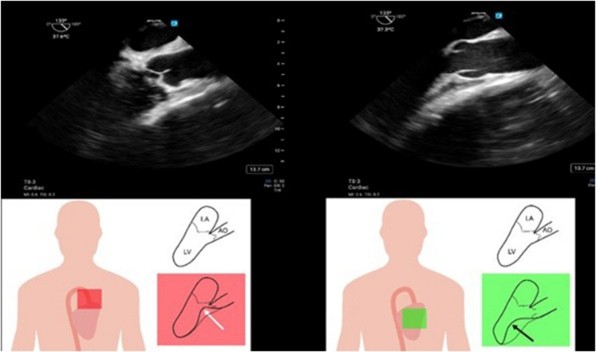
The latest issue of CASE is now available with intriguing reports, including “To Drain or Not to Drain? Pericardial Decompression Syndrome.” CASE Editor-in-Chief Vincent Sorrell, MD, FASE, remarked, “Mathias et al. help educate readers on the lesser known post-pericardiocentesis complication of pericardial decompression syndrome. They reported their findings on a complex 59-year-old patient with multiple comorbidities including acute chronic kidney disease, acidosis, hyperkalemia, atrial fibrillation treated with warfarin, pulmonary hypertension, dilated cardiomyopathy with remote ventricular tachycardia, and a large pericardial effusion with worsening hypotension. The patient had a markedly elevated international normalized ratio (INR 11) and TTE findings of tamponade. Despite urgent pericardial drainage of 600cc bloody fluid, their hemodynamic status continued to deteriorate, and the patient did not survive. The authors do an excellent job describing their thorough evaluation, approach to management, and potential underlying mechanisms, including acute pericardial decompression syndrome. Additionally, the authors provide readers with steps for when to consider this rare syndrome and how to manage this with the goal toward a different clinical outcome.”
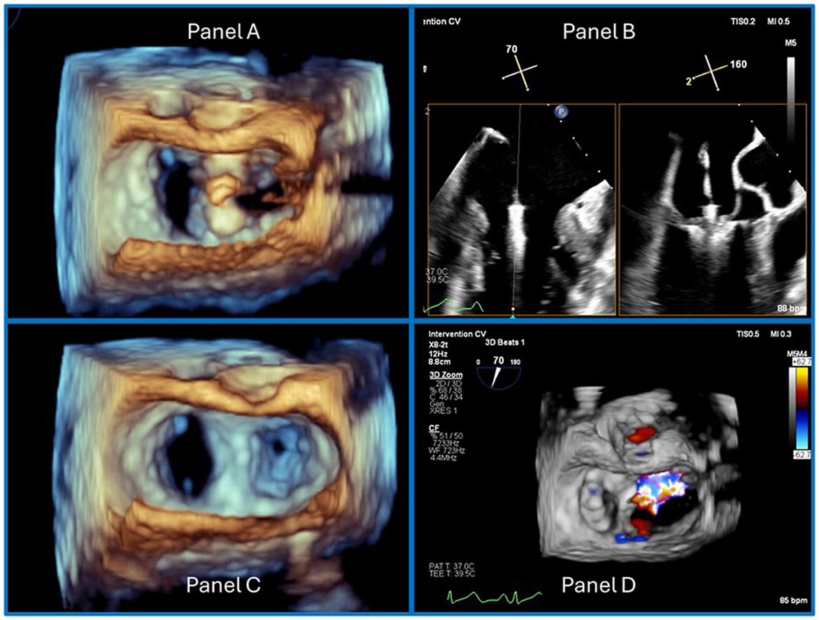
This Pericardial Pathologies report follows two cases in the Congenital Heart Disease category, including findings from a high-risk, anomalous left main coronary in a young patient with a complicated post-operative course. The other details the clinical and hemodynamic improvements of an ASD closure in an elderly patient with complex cardiovascular disease. Allow Echo Innovation to expand your sonic insights with Basit et al.’s collection of multimodality imaging findings in a case of RCA to SVC fistula. 3D TEE transillumination is used to identify a papillary fibroelastoma on the aortic valve in a highly detailed report from Gentile et al.
Contrary to the rare findings CASE often publishes, Dr. Sorrell’s editorial hones in on what it really means to mark a study as “normal.” Included is a table of 24 common cardiovascular findings that should be routinely reported in a normal study depending on the imaging modality.
Looking for a journal to submit your case report to? We want to hear from you! Email us with questions or submit your report today.
Publish date
August 20, 2024
Topic
- CASE
Related News

ASE News

Announcements

Announcements